Home » Without Label » Tendon Diagram / Feet Tendons & Ligaments | Kinesiology | Pinterest | Best ... / Tendons are thick bands of tissue that connect muscles to bones.
Tendon Diagram / Feet Tendons & Ligaments | Kinesiology | Pinterest | Best ... / Tendons are thick bands of tissue that connect muscles to bones.
Tendon Diagram / Feet Tendons & Ligaments | Kinesiology | Pinterest | Best ... / Tendons are thick bands of tissue that connect muscles to bones.. The fleshy, thick part of the muscle is called its belly. The achilles tendon is also called the calcaneal tendon. Allows the action of raising the foot. Also allows the action of raising up onto toes. Foot anatomy diagram, foot joint diagram, foot sprain diagram, foot tendons and ligaments pain, leg tendon diagram.
This sudden, tight, intense lower leg pain is sometimes called a charley horse. A tendon is a band of tissue that connects a muscle to a bone. It attaches to the wrist bone, the pisiform, and as well as the 5th hand bone. Foot anatomy diagram, foot joint diagram, foot sprain diagram, foot tendons and ligaments pain, leg tendon diagram, peroneal tendonitis, foot, foot anatomy diagram, foot joint diagram, foot sprain diagram, foot tendons and ligaments pain, leg tendon diagram, peroneal tendonitis. Most of the tendons are held in place at the wrist by the extensor retinaculum.
File:Knee diagram.svg - Wikipedia from upload.wikimedia.org The pubis, ischium, and ilium together constitute the pelvis while the thigh bone is the femur. Brings leg back to and across body. The achilles tendon enables us to walk, without it we would not be able to raise our heels of the ground. Raises and rotates arm in all directions. The achilles tendon attaches the muscles of the calves to the bones of the ankle and foot. Foot and ankle musculoskeletal key : Intermediate back muscles and c. The rotator cuff is a group of four muscles and tendons that surround the glenohumeral joint.
The achilles tendon is also called the calcaneal tendon.
Muscles of the shoulder : The achilles tendon is a tough band of fibrous tissue that connects the calf muscles to the heel bone (calcaneus). Foot anatomy diagram, foot joint diagram, foot sprain diagram, foot tendons and ligaments pain, leg tendon diagram. Flexor tendon lacerations are classified into five zones 2, 15, 16. Also allows the action of raising up onto toes. Ligaments join the knee bones and provide stability to the knee: Allows the action of raising the foot. Possibly the most important tendon in terms of mobility is the achilles tendon. It attaches to the wrist bone, the pisiform, and as well as the 5th hand bone. Extends spine and trunk back. The fcu tendon is one of two tendons that bend the wrist. The tendon is firmly connected to muscle fibres at one end and to components of the bone at its other end. Intermediate back muscles and c.
Its muscle belly is in the forearm. This sudden, tight, intense lower leg pain is sometimes called a charley horse. On the other hand, the insertion is where a tendon attaches that muscle to the *more* movable bone. The tendon travels along the inside of the forearm on the side of the small finger and crosses the wrist. Raises and rotates arm in all directions.
AEC Client Education Library - Tendon versus Ligament from www.atlantaequine.com Its muscle belly is in the forearm. Ligaments and tendons are adapted in response to changes in mechanical stiffness. The foot and ankle surgeon will select the best procedure to repair the tendon, based on the extent of the. A tendon is a band of tissue that connects a muscle to a bone. The tendon travels along the inside of the forearm on the side of the small finger and crosses the wrist. Browse 318 hand anatomy tendons stock photos and images available, or start a new search to explore more stock photos and images. The fleshy, thick part of the muscle is called its belly. For images of the muscle, click on each link under location.
Possibly the most important tendon in terms of mobility is the achilles tendon.
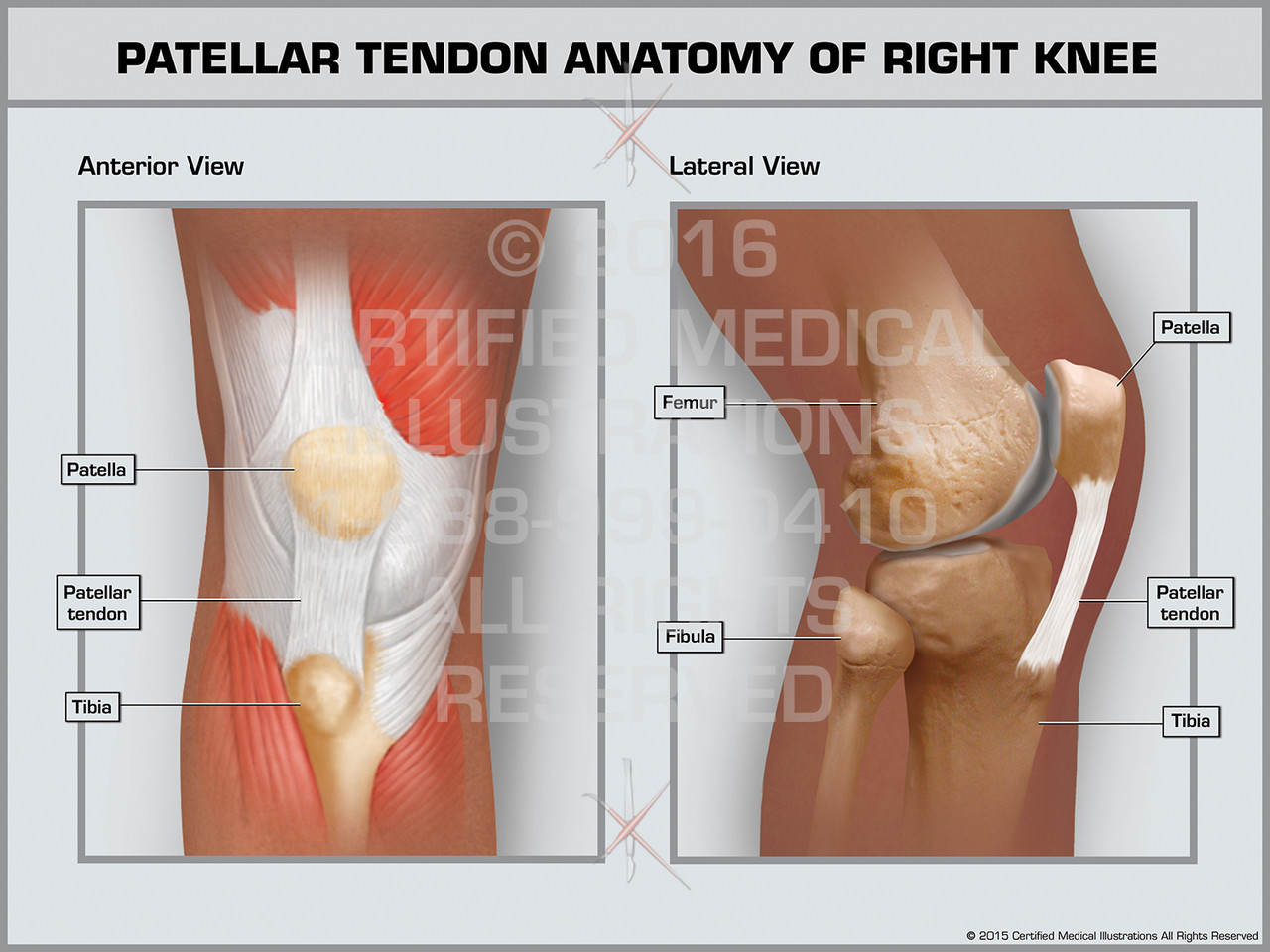
A tendon is a band of tissue that connects a muscle to a bone. This tendon connects the patella (kneecap) to the tibia. Flexes elbow and moves forearm. Tendons are thick bands of tissue that connect muscles to bones. It attaches to the wrist bone, the pisiform, and as well as the 5th hand bone. Tendons are the connection between bones and muscles. One peroneal tendon attaches to the outer part of the midfoot, while the other tendon runs under the foot and attaches near the inside of the arch. Brings leg back to and across body. Extends spine and trunk back. Tendon diagrams and design force vectors. The knee joint is a complex structure that involves bones. Foot anatomy diagram, foot joint diagram, foot sprain diagram, foot tendons and ligaments pain, leg tendon diagram, peroneal tendonitis, foot, foot anatomy diagram, foot joint diagram, foot sprain diagram, foot tendons and ligaments pain, leg tendon diagram, peroneal tendonitis. Allows the foot to be turned inward and also supports the arch of the foot.
Again, our knowledge of how mechanical stimulus mediates ligament and tendon structure is more empirical and less. Ligaments and tendons are adapted in response to changes in mechanical stiffness. Foot anatomy diagram, foot joint diagram, foot sprain diagram, foot tendons and ligaments pain, leg tendon diagram. The cause is repeated contraction of the forearm muscles that you use to straighten and raise your hand and wrist. Foot and ankle musculoskeletal key :
Patellar Tendon Anatomy of Right Knee - Print Quality ... from cdn10.bigcommerce.com This muscle diagram is interactive: Possibly the most important tendon in terms of mobility is the achilles tendon. If you tear the biceps tendon at the shoulder, you may lose some strength in your arm and have pain when you forcefully turn your arm from palm down to palm up. The achilles tendon is a tough band of fibrous tissue that connects the calf muscles to the heel bone (calcaneus). Foot anatomy diagram, foot joint diagram, foot sprain diagram, foot tendons and ligaments pain, leg tendon diagram. When the muscles tighten (contract) arguably, the most important tendon is the achilles tendon, which allows the calf muscles to move. This sudden, tight, intense lower leg pain is sometimes called a charley horse. It is controlled by the obturator nerve.
Tendons are thick bands of tissue that connect muscles to bones.
Brings leg back to and across body. This diagram depicts muscle in the body 744×1054 with parts and labels. Browse 318 hand anatomy tendons stock photos and images available, or start a new search to explore more stock photos and images. The knee joint is a complex structure that involves bones. 9 photos of the foot tendons and ligaments diagram. The tendon that attaches the biceps muscle to the forearm bones radius and. This tendon connects the patella (kneecap) to the tibia. The fcu tendon is one of two tendons that bend the wrist. Movement occurs when our muscles pull on our bones, relocating them. Black and white print showing the musculoskeletal system of a human hand, including the bones, muscles, cartilage, tendons, ligaments, and joints,. One peroneal tendon attaches to the outer part of the midfoot, while the other tendon runs under the foot and attaches near the inside of the arch. A muscle's origin is where a tendon attaches it to the *less* movable bone. Foot anatomy diagram, foot joint diagram, foot sprain diagram, foot tendons and ligaments pain, leg tendon diagram.